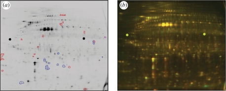
Figure 1.
DiGE gel of protein extracted from human fibroblasts (hTERT BJ-1) cultured at low density for 24 h on microgrooved (G; 5 µm deep × 25 µm pitch) versus planar (P) poly(dimethylsiloxane) (PDMS) substrates. At a 2.5-fold threshold, around 33 proteins appeared differentially regulated on this single gel. (For a multi-gel comparison, an internal standard should normally be included.) (a) Cy3 gel channel shown following software analysis with DeCyder v. 5.0. Blue, proteins upregulated in cells grown on G versus P; red, proteins downregulated in cells on G versus P. (b) Same gel as (a); green, Cy3 (extract from cells on P); red, Cy5 (extract from cells on G).

