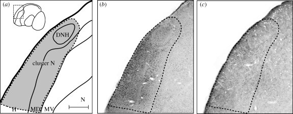
Figure 1.
ZENK activation, which mirrors neuronal activation in cluster N, is reduced to a background level when garden warblers wore hoods covering both eyes. (a) Schematic drawing of cluster N (area within dashed line). (b,c) Photos of sagittal brain slices through the centre of cluster N stained against ZENK protein. (b) ZENK activation of cluster N in a garden warbler that had both eyes open. The small black dots are ZENK-positive neuron nuclei, i.e. these neurons were active when a garden warbler had both eyes open during the night under the dim-light conditions of our wooden testing huts. (c) ZENK activation of cluster N in a garden warbler that wore one of our hoods covering both eyes. Notice that the ZENK activation level and thus the number of active neurons are reduced to the background level. Consequently, our hoods are light-tight and seem to effectively block input to the brain area cluster N, which is required for magnetic compass orientation and which seems to be involved in the processing of magnetic compass information (Zapka et al. 2009). Scale bar in (a) (for a–c) = 400 µm. DNH, dorsal nucleus of the hyperpallium; H, hyperpallium; MD, mesopallium dorsale; MV, mesopallium ventrale; N, nidopallium.

