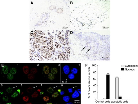
Figure 2.
Immunohistochemical detection of DBC1 in human breast tissues and colocalisation of BRCA1 and DBC1 in MCF-7 human breast cancer cells. (A–D) Breast specimens were obtained at the time of diagnosis of breast cancer in accordance with the guidelines of the Ethical Board of Komagome Hospital. DBC1 showed nuclear staining of ductal epithelium (A) and adipose tissue (B) in breast specimens. DBC1 expression was observed in the nuclei of cancer tissues (C). Cancer cells exhibiting an enlarged nucleus showed a complete loss of DBC1 expression (D, arrow). (E) MCF-7 cells were either treated or not treated by ultraviolet (UV) light (0.24 J), fixed, and permeabilised. Cells were incubated with primary antibodies and subsequently with secondary antibodies. The expression of DBC1 (red) and BRCA1 (green) was investigated under confocal fluorescence microscopy (Carl-Zeiss). Representative immunofluorescence studies are shown (E, 1–4; control, 5–8; UV exposure for 10 min, E3 and E7; merge, E4 and E8; 4′,6-diamino-2-phenylindole staining). Arrows in E5–8 indicate a cell showing apoptotic morphological changes with the cytoplasmic expression of DBC1 and BRCA1. Bars indicate 10 μm. (F) The degree of colocalisation (BRCA1 and DBC1) was measured using a confocal microscope. The colocalisation signal was quantified in the nucleus and cytoplasm of cells separately.

