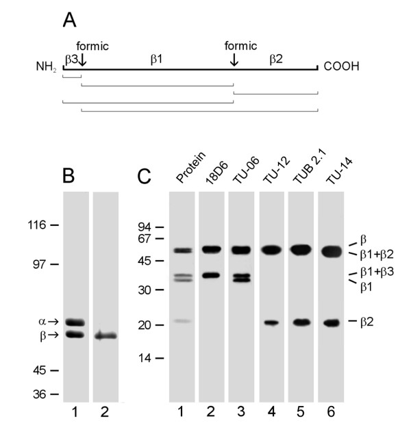
Figure 1.
Reactivity of TU-14 and TUB 2.1 antibodies with β-tubulin fragments. (A) Schematic representation of the β-tubulin peptide map after formic acid cleavage. The positions of formic acid cleavage sites (formic) and the generated proteolytic fragments (β1, β2 and β3) are shown on the thick line. Fragment sizes after incomplete cleavage are outlined below. (B) Coomassie Blue staining of carboxyamidomethylated tubulin heterodimer (lane 1) and isolated β-tubulin (lane 2). α and β denote positions of tubulin subunits. 7.5% SDS-PAGE. (C) Immunostaining with antibodies to β-tubulin fragments generated by formic acid. Lane 1: protein staining of blotted fragments; lanes 2-6: immunostaining with antibodies 18D6 (marker of β3), TU-06 (marker of β1), TU-12 (marker of β2), TUB 2.1 and TU-14. Positions of β-tubulin fragments are indicated on the right margin. 12.5% SDS-PAGE. Molecular mass markers (in kDa) are indicated on the left of B and C.

