
Figure 2.
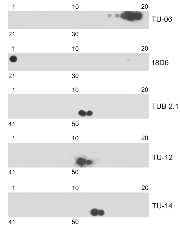
Immunostaining of peptide scans covering A. thaliana β-tubulin 1 sequences with monoclonal antibodies. Staining with antibodies 18D6 and TU-06 on peptide scans β1-180. Staining with antibodies TU-12, TUB 2.1 and TU-14 on peptide scans β171-447. Peptide scans were formed by immobilized linear 15-meric peptides with 5 amino acid overlaps. Numbers at the top and bottom denote peptide spots in the upper and lower row of the scan, respectively.