
Figure 5.
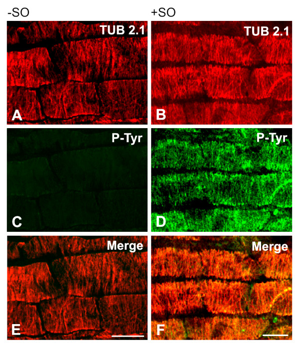
Immunofluorescence double-label staining of A. thaliana unfixed microtubules with antibodies to β-tubulin and phosphotyrosine. Preparations of primary root epidermal cells, preincubated without (A, C, E) or with (B, D, F) sodium orthovanadate (SO) to inhibit phosphatases were stained with monoclonal antibody TUB 2.1 to β-tubulin (A-B) and polyclonal anti-phosphotyrosine antibody P-Tyr (C-D). Figures A, C, E and B, D, F represent the same field. Superpositions (Merge) are shown in E and F. Bar, 10 μm.