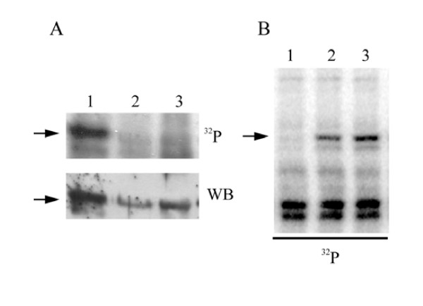
Figure 1.
Characterization of the LePRK2 dephosphorylating activity in tobacco style extracts. A, Pollen microsomal fractions (15 μg) were incubated for 10 min with [gamma-32P]-ATP in buffer without (lane 1) or with (lane 2) tobacco style extract proteins (340 μg), or with (lane 3) tomato style exudate proteins (190 μg), then separated by SDS-PAGE, blotted onto nitrocellulose and subjected to autoradiography (top panel, 32P), then incubated with anti-LePRK2 antibody (bottom panel, Western Blot). The position of LePRK2 is indicated by arrows. B, Dephosphorylation activity after chloroform extraction of style extracts. Lane 1, aqueous phase; lane 2, interface; and lane 3, organic phase. The position of LePRK2 is indicated by an arrow.

