
Figure 4.
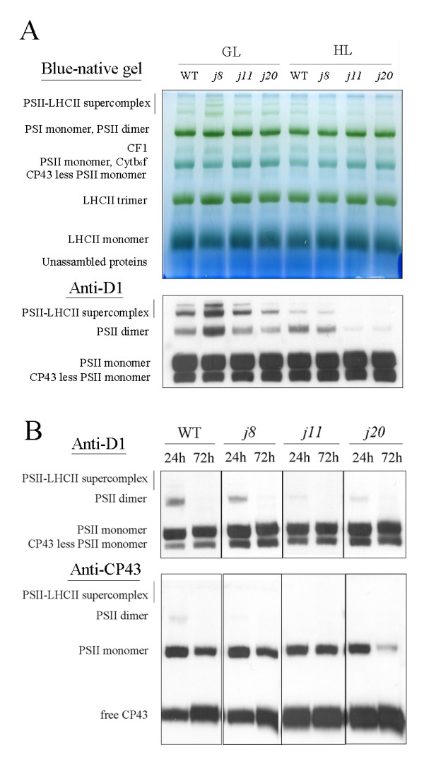
BN-PAGE analysis of thylakoid protein complexes from WT and the DnaJ mutants. Thylakoids corresponding 4 μg Chl were loaded in each lane. A, A BN gel of thylakoid protein complexes from plants exposed to growth light conditions for 6 h and from plants exposed to high light for 6 h. Top panel, BN gel directly after electrophoresis; lower panel, BN gel immunoblotted with D1 antibody. B, Immunoblots of the BN gels prepared from plants after a long-term high light (1000 μmol photons m-2 s-1) exposure. Thylakoid membrane protein complexes of WT and the DnaJ mutants were subjected to Blue-native gel electrophoresis following immunoblotting with D1 (top panel) and CP43 (lower panel) antibodies. GL, 120 μmol photons m-2 s-1 growth light; HL, 1000 μmol photons m-2 s-1 high light.