
Figure 1.
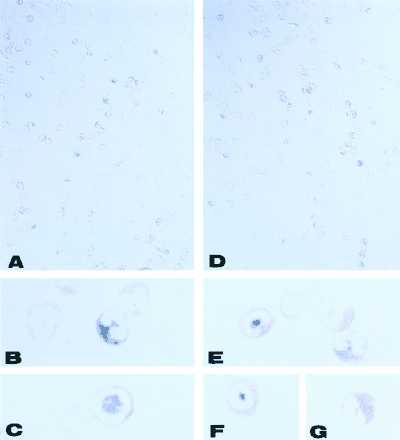
Immunolocalization of type II (A–C) and type III (D–G) NOS in cytoplasmic granules of purified rat peritoneal eosinophils. NOS was detected by using nitroblue tetrazolium/5-bromo-4-chloro-3-indolyl phosphate (blue reaction product) and counterstained with eosin. Ring-like nuclear shape (A, C, D, and F) predominated over bilobular nuclei (A, B, D, E, and G) for both NOS isotypes. Nomarski micrographs. [Original magnifications: ×33 (A and D); ×333, (B, C, E–G).]