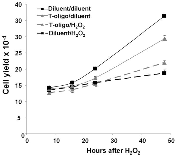
Figure 6. T-oligo protects fibroblasts from oxidative damage.
Newborn fibroblasts were processed as per Materials and Methods, and cells were then treated with 25 μM fresh H2O2 or diluent for one hour and then provided fresh medium without T-oligos. Cell yields were determined 8, 16, 24 and 48 hours later. Fibroblasts pretreated with T-oligo and then diluent proliferated more slowly than diluent pretreated cells (p< 0.0001), as expected, given the initial T-oligo induced S-phase arrest[13], but both showed exponential growth after the first 24 hours. Interestingly, fibroblasts pretreated with T-oligo and then challenged with H2O2 proliferated significantly better than diluent pretreated H2O2-challenged fibroblasts (p< 0.006). Data points are the mean ± SEM for 3 separate experiments with different donor cells.

