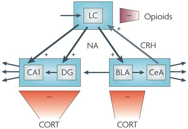
Figure 4. Direct interaction between different stress mediators.
Noradrenaline (NA), corticotropin-releasing hormone (CRH), opioids and corticosterone (CORT) interact in the locus coeruleus (LC) and its projection areas (including the hippocampus and the amygdala) to orchestrate exquisite tuning of neuronal firing patterns in response to stress. Exposure to stress shifts LC noradrenergic cell firing — which is normally kept at a moderate level by glutamatergic input, here represented by the top left arrow — from a moderate tonic activity to high tonic firing that prevents phasic firing. This shift is mediated by CRH projections from the central amygdala on to CRH 1 receptors in the LC28. In turn, LC noradrenergic cells project to the basolateral amygdala (BLA), hippocampal CA1 and the dentate gyrus (DG). Here noradrenaline, released shortly after stress exposure, enhances excitability, promoting the encoding of stress-related information. Glutamatergic output from the BLA to the DG is thought to provide a means to ‘emotionally tag’ information processed in the hippocampus, thus rendering it preference in storage71. The stress-induced enhancement in activity in the LC, the BLA, the DG and CA1 (which in the DG is aided by rapid corticosteroid actions; see main text) is gradually reversed, resulting in a return to the pre-stress activity level. In the LC, the level of tonic firing is reduced by opiates that bind to κ- and μ-opioid receptors68. In the BLA, the DG and CA1, these gradual normalizing effects are exerted by corticosterone, presumably through glucocorticoid receptor-mediated gene-dependent cascades6,70,79. The + signs indicate that the stress mediator enhances cell firing, whereas the–signs indicate decreased cell activity. CeA, central amygdala.

