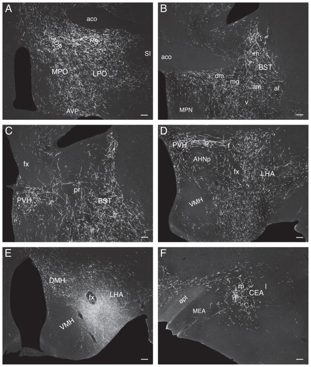
Fig. 11.
Darkfield photomicrographs showing the distribution of PHAL-labeled axons in experiment LHA22 with a PHAL injection centered caudal to the LHAsfa, in the LHAsfp. (A) Medial preoptic area, and the parastrial, anterodorsal preoptic, and anteroventral preoptic nuclei; (B) anteromedial and anterolateral areas and the rhomboid, dorsomedial, and ventral nuclei of bed nucleus of the stria terminalis’s anterior division; (C) interfascicular and transverse nuclei of bed nuclei’s the posterior division, and the paraventricular hypothalamic nucleus’s anterior parvicellular part; (D) paraventricular hypothalamic nucleus and lateral hypothalamic area; (E) dorsomedial hypothalamic nucleus and PHAL deposit centered in, but not restricted to, the LHAsfp; (F) medial part of the central amygdalar nucleus. Scale bars = 100 μm.

