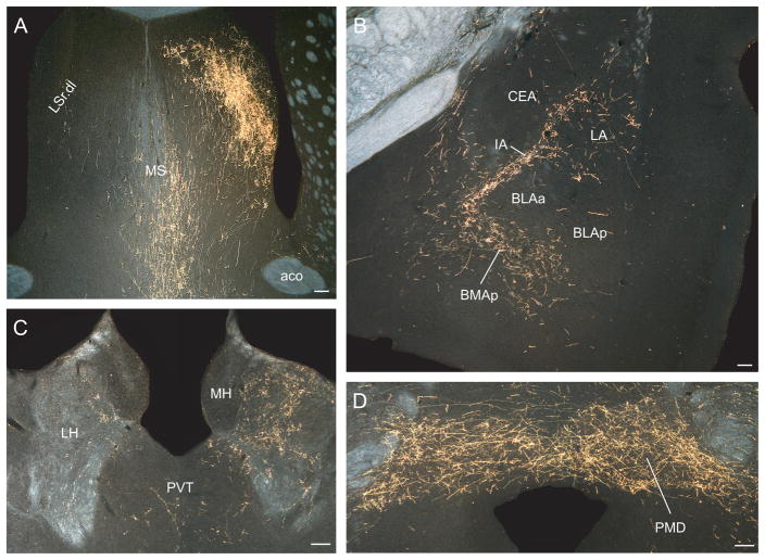
Fig. 6.
Darkfield photomicrographs showing the distribution of PHAL-labeled axons in experiment LHAsfa94, with a large injection centered in the LHAsfa. (A) Dorsolateral zone of the lateral septal nucleus’s rostral part, with an abundance of pericellular baskets; (B) intercalated and posterior basomedial nuclei of the temporal lobe’s amygdalar region; (C) lateral habenula; and (D) dorsal premammillary nucleus. Scale bars = 100 μm.

