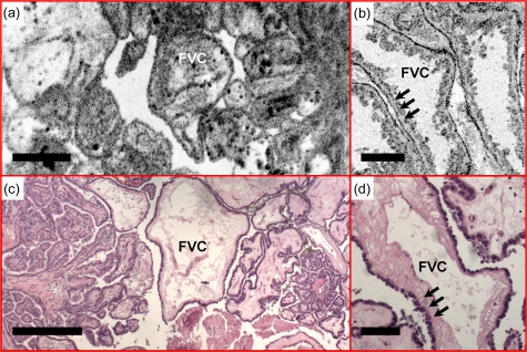
Figure 11.
Fibrovascular cores (FVCs) of the papillae, another representative feature of classic-type papillary carcinoma, are clearly identified in the (a) en face OCT and (b) OCM images obtained about 100 and 50 μm below the tissue surface, respectively. A single layer of epithelium lines the follicles (arrows), with underlying dense fibrosis. (c) and (d) are corresponding HE slides (4 and 20×, respectively). Scale bars, 500 μm in (a) and (c), and 100 μm in (b) and (d).

