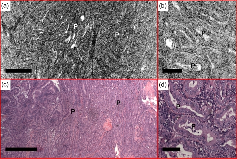
Figure 9.
Normal follicles are absent from the (a) en face OCT and (b) OCM images, obtained about 110 and 50 μm below the tissue surface, respectively, in a case of classic-type papillary carcinoma. The thyroid is replaced by complex papillae (P), showing irregular papillary fronds and complex branching features. (c) and (d) are corresponding HE slides (4 and 20×, respectively). Scale bars, 500 μm in (a) and (c), and 100 μm in (b) and (d).

