FIGURE 2.
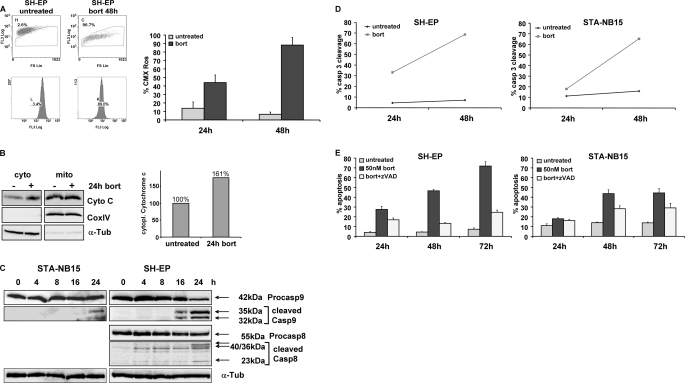
Bortezomib-induced apoptosis requires caspase activation and involves mitochondria. A, mitochondrial activity was measured by CMX-Ros staining of SH-EP cells in the presence or absence of 50 nm bortezomib for 48 h. B, cytoplasmic cytochrome c of untreated and 24 h bortezomib-treated SH-EP cells was detected by immunoblot, and the amount of cytoplasmic cytochrome c was measured by densitometry of 16-bit images using LabWorks software. C, STA-NB15 and SH-EP cells were treated with 50 nm bortezomib for 0, 4, 8, 16, and 24 h. Cell lysates were subjected to immunoblot analyses using specific antibodies against caspase-9 (with cleavage products of 35 and 32 kDa) and caspase-8 (cleavage products of 40, 36, and 23 kDa). α-Tubulin was used as loading control. D, active caspase-3 was detected by the fluorogenic FITC-DEVD.fmk substrate of the caspase-3 cleavage assay in the cells SH-EP and STA-NB15 after treatment with 50 nm bortezomib (bort) for 24 and 48 h. E, SH-EP and STA-NB15 cells were maintained for 24, 48, and 72 h in the presence of 50 nm bortezomib alone or in combination with z-VAD, a pan-caspase-inhibitor (20 nm), and subjected to PI-FACS analyses. Diagrams represent mean values of three independent experiments.

