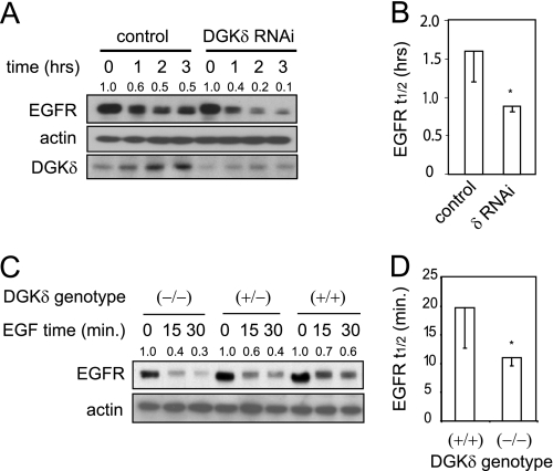
FIGURE 2.
Accelerated decay of EGFR in DGKδ-deficient cells. A, HeLa cells were transfected with scrambled or DGKδ siRNA duplexes, starved for 16 h, and then treated with EGF (5 ng/ml) for 0–3 h. EGFR, actin, and DGKδ were then detected in cell lysates by Western blotting. EGFR band densities (arbitrary units) are shown above the blot. Similar results were obtained when cyclohexamide (5 μm) was included to minimize new protein synthesis. B, the half-life (t½) of EGFR was measured in three experiments like that shown in A. Shown are mean values with S.D. (*, p < 0.05). C, EGFR decay in primary keratinocytes from wild-type mice or mice with heterozygous or homozygous deletion of DGKδ was measured after treatment with EGF (10 ng/ml) for the indicated times. EGFR band densities (arbitrary units) are shown above the blot. D, the half-life (t½) of EGFR was measured in three experiments like that shown in C. Shown are mean values with S.D. (*, p < 0.05).

