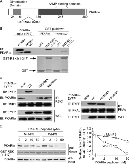
FIGURE 2.
RSK1 binds to the pseudosubstrate region of PKARIα. A, shown is a schematic of bovine PKARIα; numbers represent the amino acids in bovine PKARIα. B, the truncated PKARIα (deletion of amino acids 1–91, PKARIαΔ91) retains the ability to interact with RSK1. Pure PKARIα or PKARIαΔ91 (10 pmol each) was incubated with glutathione-Sepharose prebound with GST or GST-RSK1-(1–317) (5 μg). The amounts of PKARIα or PKARIαΔ91 in the pulldown complex were detected with anti-PKARIα antibody. GST or GST-RSK1-(1–317) was stained with Coomassie Blue. IB, immunoblot. C, substitution of Arg-93/94 or Arg-95/96 on PKARIα to Ala selectively abrogates the interactions of PKARIα with RSK1 and PKAc. HEK293T cells were transfected with plasmids expressing C-terminal fusion of Wt-PKARIα, PKARIα (R93A/R94A), or PKARIα (R95A/R96A) with enhanced yellow fluorescent protein (wt-PKARIα-EYFP, PKARIα (R93A/R94A)-EYFP, and PKARIα (R95A/R96A)-EYFP, respectively). Cell lysates were immunoprecipitated (IP) with anti-RSK1 or PKAc antibody. The immune complex was probed with anti-EYFP, PKARIα, RSK1, or PKAc antibodies. WCL, whole cell lysate. D, the peptide corresponding to the PKARIα pseudosubstrate region (Wt-PS) competes for the interaction of PKARIα with RSK1. GST-RSK1-(1–317) (5 μg) prebound to glutathione-Sepharose was incubated with PKARIα peptides, Wt-PS, or Mut-PS at the concentrations indicated at 4 °C for 15 min before being mixed with PKARIα (10 pmol). GST-RSK1-(1–317) was stained with Coomassie Blue. The panel on the right shows the quantification of relative intensities of PKARIα bands as a ratio of GST-RSK1 bands from two identical experiments. Wt-PS, peptide with wild-type PKARIα pseudosubstrate sequence KGRRRRGAI). Mut-PS, peptide with mutated PKARIα pseudosubstrate sequence (KGAARRGAI).

