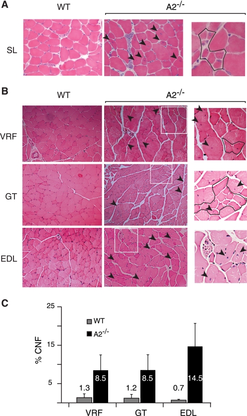
FIGURE 7.
Histological evidence of myopathy in 8–10-month-old APOBEC2−/− mice. A, examples of a region of soleus muscle (SL) in 15-week-old wild-type (WT) or APOBEC2-deficient (A2−/−) mice. The arrowheads indicate fibers with centrally located nuclei, and the right panel shows a region with immature myotubes. B, comparison of transverse sections of vastus and rectus femoris (VRL), gastrocnemius (GT), and EDL muscle from 9-month-old APOBEC2-deficient and wild-type mice. The boxed areas in the middle panels are shown in higher magnification in the right panels, where outlined regions contain smaller fibers. C, histogram comparing the percentage of centrally nucleated fibers (CNF) in different muscles from 9-month-old APOBEC2-deficient and control mice.

