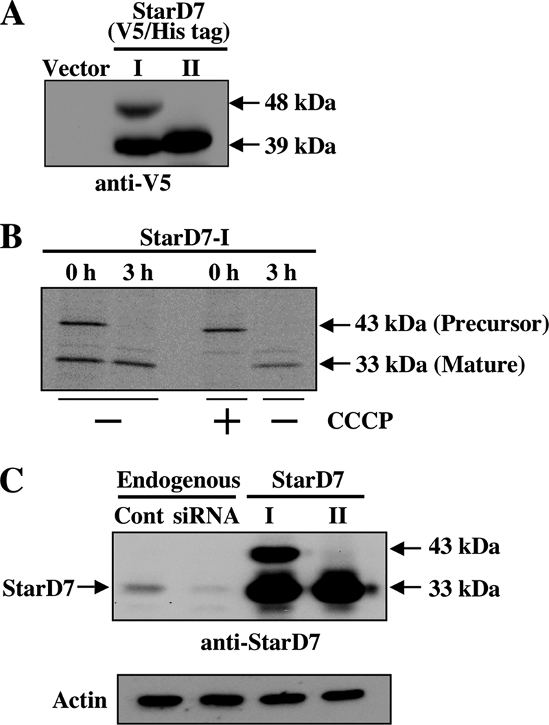
FIGURE 2.

Proteolytic processing of StarD7-I. A, V5/His-tagged StarD7-I and -II were expressed in HEPA-1 cells (60–70% confluent), and cell lysates were analyzed by Western blotting with anti-V5 antibody. B, pulse-chase experiment. StarD7-I without tag was overexpressed in HEPA-1 cells, and proteins were pulse-labeled with 30 μCi/ml of [35S]Met and [35S]Cys for 20 min with or without CCCP. Then, cells were cultured in normal medium for 3 h. Proteins were immunoprecipitated with anti-StarD7 antibody, and precipitated proteins were separated by SDS-PAGE. C, molecular mass of endogenous StarD7. HEPA-1 cells were transfected with the StarD7-specific siRNA or the expression vector for StarD7-I or StarD7-II, and cell lysates were analyzed by Western blotting with anti-StarD7 antibody.
