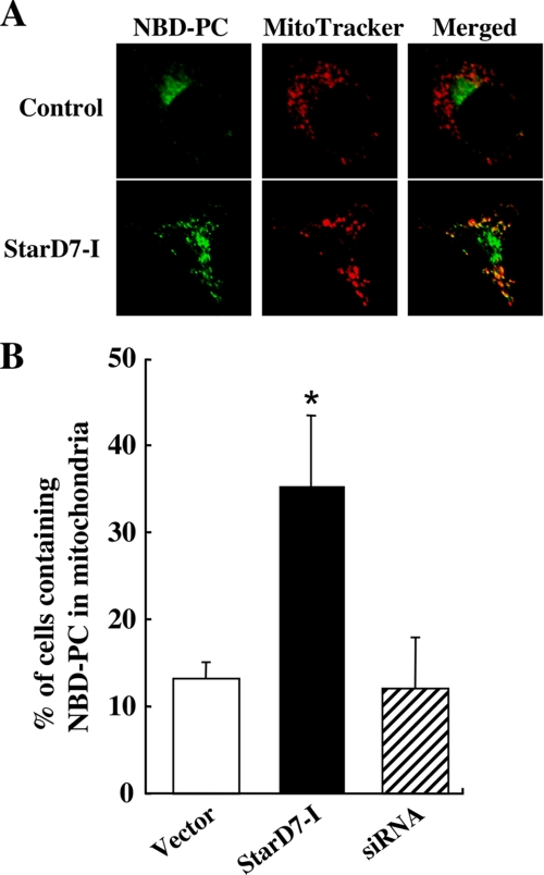
FIGURE 5.
Intracellular transport of fluorescent PC analog in cells that overexpress StarD7-I. A, intracellular localization of fluorescent PC exogenously incorporated into HEPA-1 cells. Cells (60–70% confluent) transfected with empty vector or the expression vector for StarD7-I were incubated with lipid vesicles containing C6-NBD-PC (green) and then with MitoTracker Red (red). Cells were fixed and analyzed by confocal microscopy. Yellow indicates the co-localization of the green and red signals. B, quantification of cells showing the co-localization of NBD-PC and MitoTracker Red. Cells (60–70% confluent) transfected with empty vector, the expression vector for StarD7-I, or StarD7-specific siRNA were incubated with NBD-PC and MitoTracker. Values are means ± S.D. from four independent culture dishes. *, p < 0.01 as compared with the vector control.

