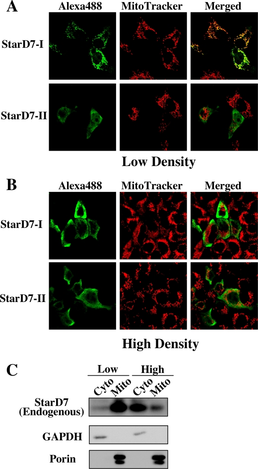
FIGURE 6.
Intracellular localization of StarD7-I and -II. HEPA-1 cells plated at low density (20–30% confluent) (A) or high density (100% confluent) (B) were transfected with the expression vector for V5/His-tagged StarD7-I or -II, and then immunostained with anti-V5 antibody followed by anti-mouse IgG Alexa488 (green) and MitoTracker Red (red). Yellow indicates the co-localization of their signals. C, subcellular fractionations of HEPA-1 cells cultured at different cellular densities. Mitochondrial and cytoplasmic fractions were prepared from cells cultured at low density (20–30% confluent) or high density (100% confluent) and analyzed by Western blotting with anti-StarD7 antibody. The purity of the mitochondrial or cytosolic fraction was verified by anti-porin or anti-GAPDH antibody.

