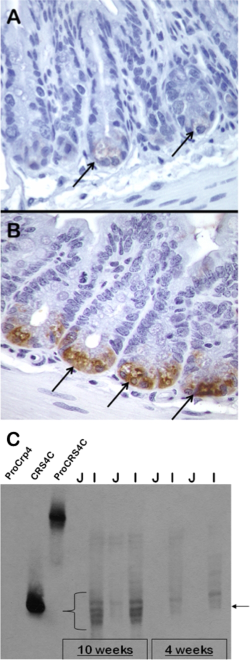
FIGURE 2.
Temporal and regional CRS4C peptide levels in SAMP1/YitFc mouse small intestine. A and B, immunohistochemical staining of crypts of the non-ileitis-prone AKR (A) and SAMP1/YitFc (B) mouse strains using anti-CRS4C-1 antiserum (see “Experimental Procedures”). Arrows indicate strongly immunopositive SAMP1/YitFc Paneth cells in B and AKR Paneth cells in A that exhibit markedly lower levels of immunoreactivity. CRS4C peptides are products of Paneth cells in the intestinal crypts of both mice. C, age-related appearance and regional distribution of CRS4C peptide accumulation determined for the SAMP1/YitFc mouse by Western blotting. Samples (0.50 mg) of total organ protein-extracted jejunum (J) and ileum (I) of individual 4- or 10-week-old SAMP1/YitFc mice were separated by AU-PAGE as noted and blotted onto a nitrocellulose membrane. The blot was subjected to Western blot analysis using anti-CRS4C-1 IgG diluted 1:5 (see “Experimental Procedures”). Recombinant pro-Crp-4 (leftmost lane) was a negative control peptide, and recombinant CRS4C-1 and pro-CRS4C-1 provided positive controls as shown by their strong immunoreactivity. Immunopositive bands that co-migrate with recombinant CRS4C-1, indicated by the lower right arrow and the curly brace, are readily detected in ileum of both 4- and 10-week-old SAMP1/YitFc mice, with the intensity of the signal markedly increased by 10 weeks of age.

