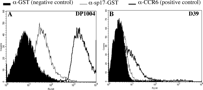
FIGURE 2.
Presence of PfbB on the bacterial surface as assessed by immunofluorescence flow cytometry analysis of the unencapsulated DP1004 strain (A) and of the encapsulated D39 strain (B). Bacteria were exposed to mouse antisera raised against GST (α-GST, used as a negative control), against a choline-binding protein-enriched fraction (α-CCR6, used as a positive control), or against the sp17 fragment of PfbB fused to GST (sp17-GST). Antibody binding was detected with phycoerythrin-conjugated goat anti-mouse IgG.

