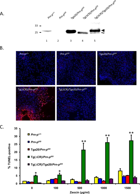
FIGURE 5.
Differentiated NSCs expressing ΔCR PrP are hypersensitive to drugs. A, PrP expression in differentiated NSC clones of the indicated genotypes was analyzed by Western blotting using 8H4 antibody. Samples were enzymatically deglycosylated with PNGase F. Bands corresponding to WT and ΔCR PrP are indicated by black and white arrowheads, respectively. B, NSCs dissected at embryonic day 13.5 mouse embryos of the indicated genotypes were cultured as neurospheres and differentiated for 7 days in the presence of retinoic acid. Differentiated NSCs were treated for 72 h with Zeocin (500 μg/ml) and were then stained by TUNEL (red) to reveal fragmented DNA and with DAPI (blue) to reveal nuclei. C, differentiated NSCs (two independent clones for each genotype) were treated for 72 h with the indicated concentrations of Zeocin. The number of TUNEL-positive cells, expressed as a percentage of the number of DAPI-stained cells, was determined in five fields for each sample group. The bars show means ± S.E. (n = 3 independent experiments). The number of TUNEL-positive cells was significantly higher in Tg(ΔCR)/Prn-p0/0 cells than in control cells at all drug concentrations (*, p < 0.05; **, p < 0.01).

