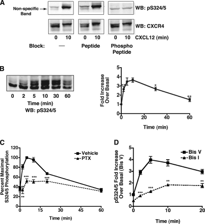
FIGURE 3.
Characterization of anti-Ser(P)-324/5 (pS324/5). A, shown is a representative Western blot (WB) demonstrating the specificity of the Ser(P)-324/5 antibody. Ten μg of purified antibody was incubated for 10 min with vehicle (PBS), 10 μg of peptide (C-Ahx-RGSSLKIL), or 10 μg of phosphopeptide (C-Ahx-RG(pS)(pS)LKIL) before overnight incubation with the nitrocellulose blots. B, cells stably expressing FLAG CXCR4 were stimulated at the time points indicated with 100 nm CXCL12. Lysates were processed and separated to visualize the agonist promoted gel shift of CXCR4. Blots were incubated overnight with a 1:1000 dilution of crude Ser(P)-324/5 antibody. Ser(P)-324/5 was normalized to total CXCR4, and data are presented as the -fold increase over basal (±S.E., n = 4). The -fold increase at 10 min is significantly different from 2, 30, and 60 min (p = 0.004, 0.05, and 0.009, respectively) but not 5 min. C, cells stably expressing FLAG CXCR4 were treated overnight with vehicle (PBS) or pertussis toxin (PTX, 100 ng/ml) before stimulation with 100 nm CXCL12. Ser(P)-324/5 was normalized to total CXCR4, and data are presented as the percent maximum of vehicle-treated cells (± S.E., n = 3). D, cells stably expressing FLAG CXCR4 were serum-starved for 6 h. 30 min before CXCL12 stimulation cells were pretreated with 2.5 μm Bis I or Bis V. Ser(P)-324/5 was normalized to total CXCR4, and data are presented as the -fold increase over basal (Bis V) (±S.E., n = 4; *, p ≤ 0.05; **, p ≤ 0.01; ***, p ≤ 0.001).

