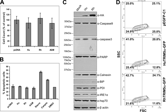
FIGURE 9.
Overexpression of TRIM5α does not induce an apoptosis. A, HEK293T cells were transfected with HA-tagged human, rhesus, or AGM TRIM5α, or an empty vector pcDNA3.1, and 48 h later the number of live cells was counted after trypan blue staining. The values are shown as % of the control. Data are expressed as the mean ± S.D. of three independent experiments. B, HEK293T cells were transfected with HA-tagged human, rhesus, or AGM TRIM5α, or an empty vector pcDNA3.1, and 48 h later the percentage of apoptotic cells were determined by using the Annexin V-FITC Apoptosis Detection Kit I (BD Biosciences). When indicated, HEK293T cells were treated with staurosporine at 10 μm for 18 h or with camptothecin at 12 μm for 5 h to induce apoptosis. Data are expressed as the mean ± S.D. of three independent experiments. C, HEK293T cells were transfected with HA-tagged human or rhesus TRIM5α, or an empty vector pcDNA3.1, and 48 h later the cell lysates were subjected to Western blot analysis to detect HA-TRIM5α proteins, apoptosis marker proteins (caspase-9, caspase-3, and PARP), ER stress marker proteins (calnexin, BiP, PDI, and IRE1α), Hsp70, or β-actin as an internal control. D, HEK293T cells were transfected with pEGFP-C1, human TRIM5α-GFP, or rhesus TRIM5α-GFP, and 48 h later the forward scatter (FSC) and side scatter (SSC) patterns of GFP-positive cells were analyzed by a flow cytometer: Q1 (low FSC and high SSC), Q2 (high FSC and high SSC), Q3 (low FSC and low SSC), and Q4 (high FSC and low SSC). Data shown here is one representative data of three independent experiments.

