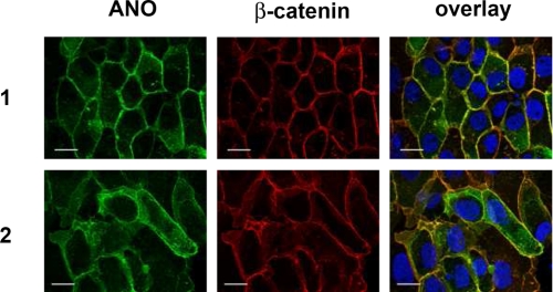
FIGURE 3.
ANO1 and ANO2 are associated with the plasma membrane of FRT cells. His-tagged ANO1 and ANO2 were overexpressed in FRT cells and were detected with anti-His antibody. β-Catenin was visualized using an Alexa 568-labeled phalloidin. Plasma membrane staining of ANO1 and ANO2 is shown by colocalization with β-catenin (yellow color in the overlay picture). No immunofluorescence was detected in mock-transfected control cells.

