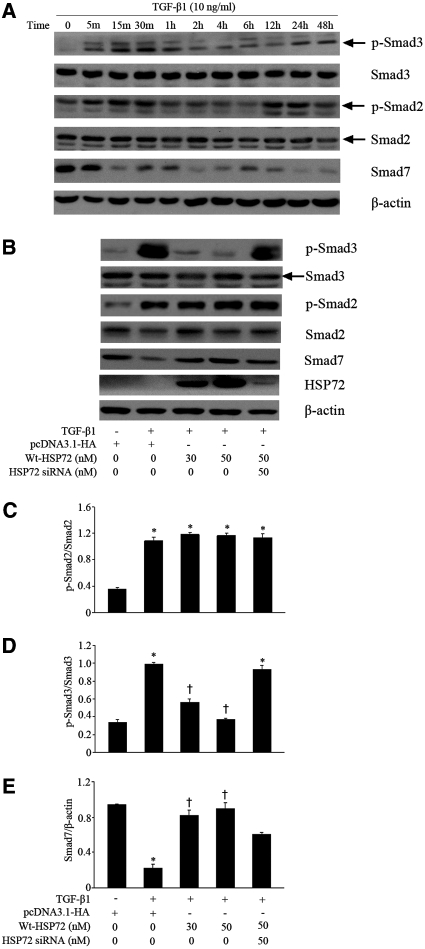
Figure 3.
HSP72 suppresses activation of the TGF-β/Smads pathway. (A) Serum-deprived NRK-52E cells were treated with 10 ng/ml of TGF-β1 for the indicated time period. Cell lysates were probed with antibodies against p-Smad3, Smad3, p-Smad2, Smad2, Smad7 or β-actin. (B) NRK-52E cells were transfected with pcDNA3.1-HA-Wt-HSP72 (30 and 50 nM) or specific HSP72 siRNA were stimulated with 10 ng/ml of TGF-β1 for 30 minutes. (C through E) Smad protein content evaluated by Western blotting with densitometric analysis of the effect of HSP72 expression on p-Smad3, p-Smad2 and Smad7 content normalized with Smad2, Smad3, or β-actin content in TGF-β1-treated cells. Data are expressed as mean ± SEM; n = 3 per treatment; *P < 0.01 versus negative control; †P < 0.05 versus TGF-β1-treated cells without HSP72 overexpression or HSP72 siRNA.

