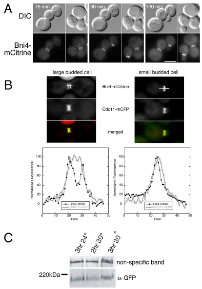
Fig. 2.
Bni4-mCitrine localization in large budded cells. (A) Time-lapse imaging of DIC and fluorescence images of cells from strain KT2575 (BNI4-mCitrine cdc13-1) after a shift from 24°C to 32°C. The time indicated in each panel is the time after the temperature shift. Note the small budded cells at the 90 minute and 120 minute time points still retain Bni4-mCitrine on the daughter-side of the bud neck. (B) Upper panels: fluorescence images of septin (Cdc11-mCFP) and Bni4-mCitrine in diploid strain JRL265/JRL574 (BNI4-mCitrine homozygous CDC11-mCFP heterozygous). Cells were grown to mid-log phase in synthetic medium and then incubated for 1.5 hours in 150 mM hydroxyurea. Lower panels: the relative fluorescence of the CFP and mCitrine was measured through the middle of the bud neck along the mother-bud axis for 3 μm as illustrated by the bars in the upper panels. (C) Immunoblot analysis of Bni4-mCitrine using α-GFP. Strain KT2575 (BNI4-mCitrine cdc13-1) was grown to mid-log phase in YPD at 24°C and then incubated for the indicated times at either 24°C or 30°C. A non-specific band was used as a load control.

