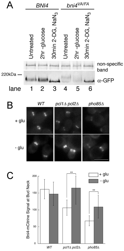
Fig. 7.
Bni4 and Bni4V831A/F833A become hypophosphorylated following energy depletion. (A) Immunoblot of Bni4-GFP and Bni4V831A/F833A-GFP using α-GFP. Strains JRL369 (BNI4-GFP) (lanes 1-3) and YLK85 (bni4V831A/F833A-GFP) (lanes 4-6) were grown to mid-log phase in synthetic medium containing glucose. Cells were then left untreated (lanes 1 and 4) or incubated in synthetic media lacking glucose (lanes 2 and 5) with the addition of 100 mM 2-deoxyglucose and 10 mM NaN3 (lanes 3 and 6) for the indicated times. A non-specific band was used as a load control. (B) Fluorescence microscopy of Bni4-mCitrine at the neck in wild-type (JRL265), pcl1Δ pcl2Δ (JRL685) and pho85Δ (JRL653) cells. Cells were imaged at mid-log phase in synthetic medium (+ glu) or after incubation for 1.5 hours in synthetic medium without glucose (− glu). Scale bar: 5 μm. (C) Quantitative analysis of relative fluorescent signal at the bud neck of the cells in B (n>50). Double asterisks denote significantly different levels. **P<0.0001, according to the unpaired Student's t-test.

