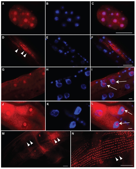
Fig. 3.
Immunostaining identifies endogenous FRG-1 as both a nuclear and a cytoplasmic protein displaying a pattern indicative of body-wall muscle dense bodies. N2 worms immunostained for FRG-1 (A,D,G,J,M,N) in red and co-stained with DAPI (B,E,H,K) in blue, with images merged (C,F,I,L). In embryo (A-C), larval (D-I) and adult (J-N) stages, FRG-1 was localized in the DAPI-poor regions of the nuclei (I and L, arrows). Cytoplasmic staining for FRG-1 resembled that of body-wall muscle cell dense bodies (D,M,N, arrowheads). Scale bars: 10 μm.

