Fig. 6.
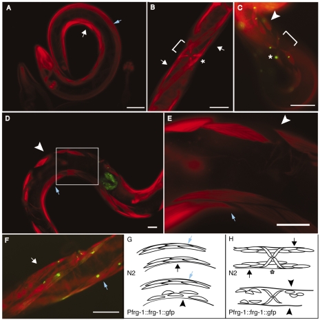
Overexpression of FRG-1 specifically from the frg-1 promoter disrupts the ventral muscle-muscle lateral junctions. Phalloidin staining of (A) wild-type N2 adults and (B) pRF4(rol-6) transgenic animals shows the normal organization and structure of the ventral (white arrows) and dorsal (blue arrows) body-wall and vulval (asterisk) muscles. Transgenic animals overexpressing FRG-1::GFP from the frg-1 promoter visualized by phalloidin and GFP merge (C-E) show specific disruption of ventral muscle-muscle lateral junctions and absence of some muscle cells (white arrowheads, bracket at vulva), whereas the dorsal musculature (blue arrows) remains intact. The boxed area in D is shown magnified 3× in E. (F) Transgenic animals overexpressing FRG-1::GFP from the myo-3 promoter showed normal muscle structure, as visualized by phalloidin staining. Diagrams depicting a lateral view (G) and a ventral view (H) of the dorsal (blue arrows) and ventral (black arrows, normal; black arrowheads, disrupted) musculature of normal (top) and transgenic (bottom) animals. Scale bars: 50 μm.

