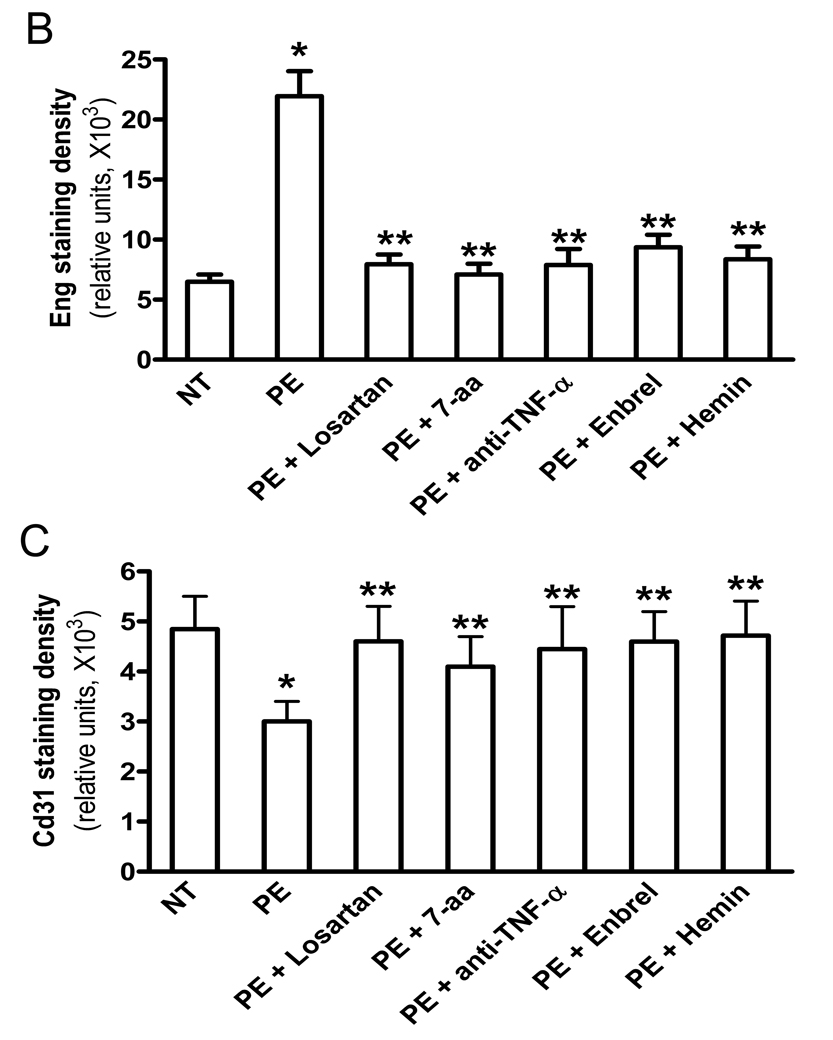
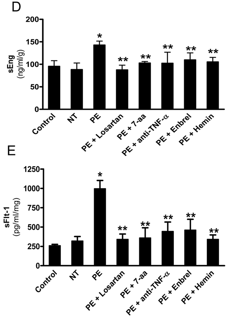
Figure 5. AT1-AA-mediated TNF-α induction contributes to impaired placenta angiogenesis by inducing both sEng and sFlt-1 secretion from human villous placental explants.
Normal human placental villous explants were collected and treated with IgG from women with preeclampsia or normotensive pregnant individuals in the presence or absence of various reagents for 72 hours. (A) At the end of treatment, human placental villous explants were collected, fixed and stained with (H&E), anti-human Eng and anti-human CD31 antibody. Scale bar, 100 µm (H&E and CD31) or 200 µm (Eng). Syncytiotrophoblast (syn) is indicated by arrow head. (B–C) Expression of endoglin (B) and CD31 (D) were quantified using Image-Pro Plus image analysis sosftware. (E–F) Cell culture supernatants were collected for sEng (E) and sFlt-1 (F) measurements by ELISA. Data are expressed as mean ± SEM of ≥4 experiments performed in duplicate (n=12–14 patient’s IgG for each category). * P < 0.001 versus villous explants treated with normotensive IgG. ** P < 0.05 versus villi treated with preeclamptic IgG.


