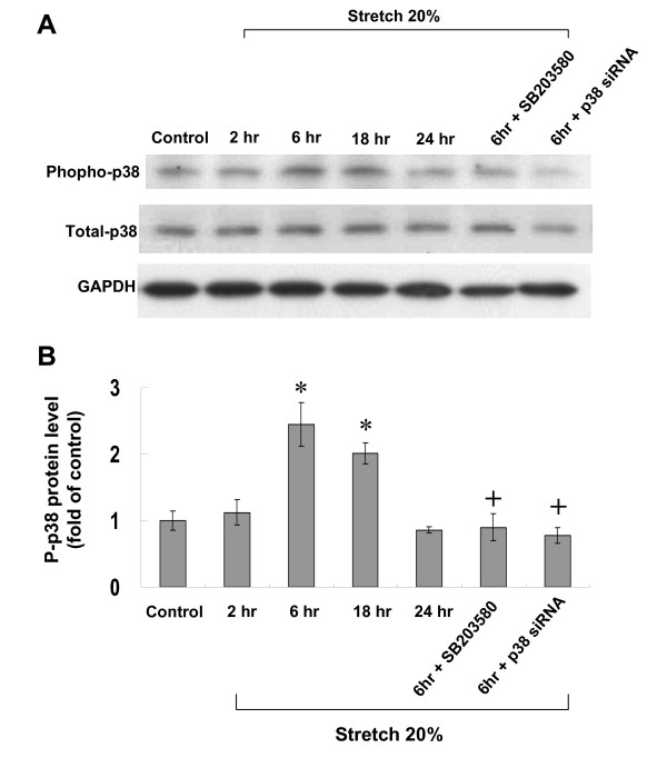
Figure 3.
Effect of cyclic stretch on expression of p38 kinase in cardiomyocytes. (A) Representative Western blots for phosphorylated and total p38 kinases in cardiomyocytes after stretch by 20% for various periods of time and in the presence of SB203580 and p38 siRNA. (B) Quantitative analysis of phosphorylated p38 protein levels. The values from stretched cardiomyocytes have been normalized to matched GAPDH and corresponding total protein measurement and then expressed as a ratio of normalized values to each phosphorylated protein in control cells. Data are from 4 independent experiments. *P < 0.001 vs. control. +P < 0.001 vs. stretch 6 h.

