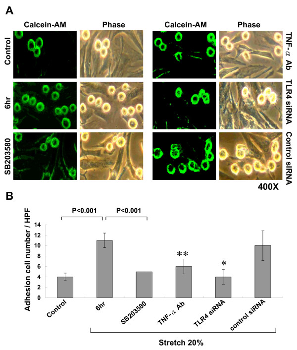
Figure 8.
Cyclic stretch increases adhesion of monocyte to stretched cardiomyocytes. A, Representative microscopic image for monocyte adhesion assay with (left panel) or without green fluorescence (right panel) in cardiomyocytes subjected to cyclic stretch for 6 h or control cells without stretch in the absence or presence of inhibitors. B, Quantitative analysis of the positive fluorescent cells. (n = 4 per group). *P < 0.001 vs. 6 hr. **P < 0.01 vs. 6 hr

