Figure 3.
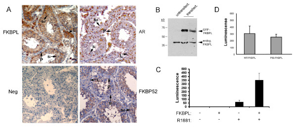
FKBPL expression in human testis. A. Immunohistochemical staining of human testis sections with the indicated antibodies. AR shows nuclear staining in Sertoli (Si), Leydig (L) and peritubular myoid (P) cells, but is negative elsewhere, including spermatids (Sd). FKBP52 is cytoplasmic, staining Sertoli and spermatogenic cells, but shows weak or absent staining in Leydig and myoid cells. FKBPL is also cytoplasmic, matching FKBP52 in most respects except for strongly staining the Leydig cells. Preimmune serum for FKBPL (shown) and other antibodies was used as a negative control (neg.). Experiments were repeated three times. Staining for FKBPL and FKBP52 was almost identical in rat testis (not shown). B. Western blot of total protein from untransfected HT29 cells (untransfect.) or transfected with a GFP-FKBPL fusion protein (transfect.). The size of the endogenous protein (endog.) is indicated. The experiment was repeated three times. C. LNCaP cells were transfected with a construct (pPA) containing luciferase driven by the prostate specific antigen transcriptional regulatory elements either with or without FKBPL as shown. The indicated cultures were additionally exposed to 1 nM of AR ligand (R1881). Luciferase activity was measured using a fluorimeter and normalised to an internal control. The experiment was carried out more than three times with similar results. D. LnCaP cells were transfected with pPA and FKBPL from normal controls (WT) or from the mutated allele seen in patient 83 (p83- listed as #6 in Table 1) and exposed to R1881. The results shown represent the median of three experiments.

