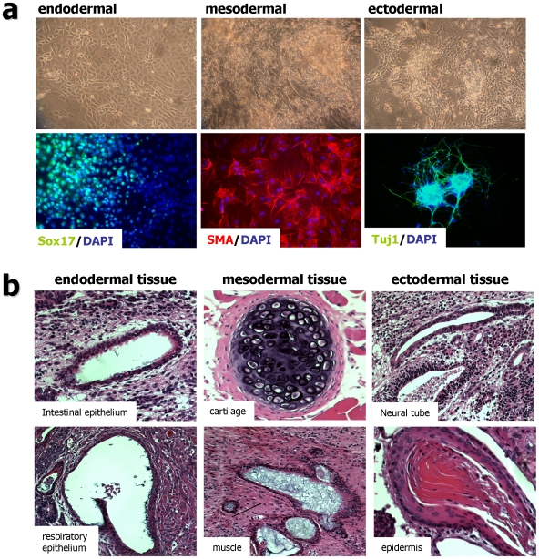
Figure 3. In-vitro and in-vivo differentiation of rat fibroblast derived iPS cells (REF-iPS #2 and #3).
(a) Embryoid body (EB) mediated in vitro differentiation of rat iPS clones. Upper panel show phase contrast images of differentiated cells at 10 days after EB attachment. Definitive endoderm-like (left), contracting muscle-like (middle) and neuronal-like (right) cells are shown. Immunocytochemical analysis for the three germ layer differentiation was performed 10 days after EB attachment (Lower images). Sox17 (green, endodemal; left), smooth muscle actin (SMA, red, mesodermal; middle) and Tuj1 (green, ectodermal; right). Nuclei were stained with DAPI (blue). (b) Hematoxylin and eosin staining of teratoma derived from rFC-iPS cells (#2 and #3). Cells were transplanted into the kidney capsule of three SCID mice. A tumor developed from one injection site. Images are from a teratoma containing intestinal epithelium, respiratory epithelium (both endodermal); cartilage, muscle (both mesodermal); neural tube, epidermis (both ectodermal).

