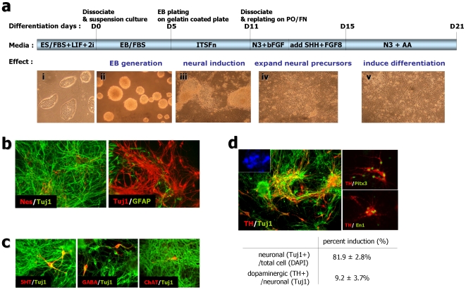
Figure 4. Rat fibroblast derived-iPS cells (REF-iPS#2 cells) progressively induced into dopaminergic neurons.
(a) Scheme of the rat iPS differentiation method used to induce dopaminergic neurons from rat iPS cells. Bottom pictures (i∼v) show representative image of each stage. (b) The five-stage neuronal differentiation method induced diverse neural differentiation in rat iPS cells. Six days after withdrawal of the mitogen bFGF at day 21 of the differentiation procedure, expression of nestin (b, left, red), Tuj1 (b, left, green; b, right, red) and GFAP (b, right, green) confirmed the neural identity of the REF-iPS derived neural differentiation. (c) Diverse subtypic neurons are expressed during rat iPS derived neuronal induction. Serotonergic (5-HT, red, left), GABAnergic (GABA, red, middle) or cholinergic (ChAT, red, right)/Tuj1 (green)-positive neurons were induced. Images were taken from REF-iPS#2 derived neuronal cells at day 21. (d) Dopaminergic neuronal differentiation from rat iPS#2-derived neuronal induction at day 21. Representative images of TH (red)/Tuj1 (green)-positive neurons derived from rat iPS cells (Inset, DAPI nuclear staining of the same field). The bottom table shows dopaminergic neuronal differentiation efficiency of rat fibroblast derived iPS cells (REF-iPS #2; from nine independent experiments, mean ± S.E). Indicated are the percent of neuronal (Tuj1+) cells per total (DAPI) cells or dopaminergic (TH+) cells per neurons (Tuj1+) at neuronal induction day 21.

