Figure 3.

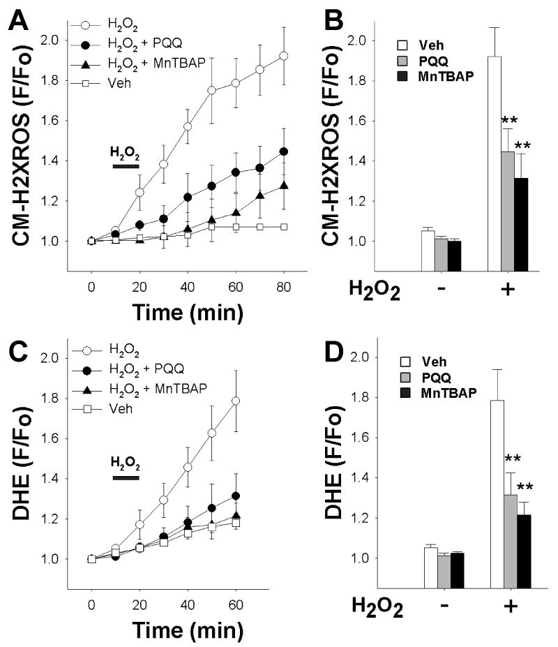
PQQ protects against an increase in cellular reactive oxygen species (ROS) in acutely isolated adult rat cardiac myocytes.
(A) Rat cardiomyocytes were loaded with CM-H2XRos to detect the increase in intracellular levels of ROS. CM-H2XRos fluorescence was collected, and fluorescence (F) at the indicated timepoint was normalized to baseline fluorescence (Fo) and plotted over time. Bar indicates addition of H2O2. Each point represents the mean ± SEM of 3-5 coverslips, each prepared from a separate rat heart. For each coverslip, 1-3 cells were analyzed. Veh – vehicle treatment (control); H2O2 (1 mM); PQQ (10 μM); MnTBAP (10 μM); PQQ and MnTBAP were administered for at least 30 minutes prior to H2O2.
(B) Peak cellular levels of ROS detected as a function of CM-H2XRos normalized to baseline value after 60 min H2O2 exposure. PQQ (gray bars) and MnTBAP (black bars) significantly reduced fluorescence compared to control (white bars).
(C) Cardiomyocytes were loaded with DHE to detect the increase in intracellular levels of ROS. DHE fluorescence was collected, and fluorescence (F) at the indicated timepoint was normalized to baseline fluorescence (Fo) and plotted over time. Arrow indicates addition of H2O2. Each point represents the mean ± SEM of 3-5 coverslips, each derived from a separate rat heart. For each coverslip, 1-3 cells were analyzed. Veh – vehicle treatment (control); H2O2 (1 mM); MnTBAP (10 μM); PQQ and MnTBAP were administered for at least 30 minutes prior to H2O2.
(D) Peak cellular levels of ROS detected as a function of DHE normalized to baseline value after 40 min H2O2 exposure. PQQ (gray bars) and MnTBAP (black bars) significantly reduced fluorescence compared to control (white bars). ** p<0.01 compared to control (H2O2 alone).
