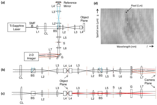
Fig. 1.
(a) Schematic of line-field phase microscope (LFPM). (b,c) Horizontal and vertical perspectives, respectively, of the LFPM. (d) Typical 2-D recorded interferogram illustrating spectral and spatial measurements along the two orthogonal directions of the 2-D spectrometer. SMF: single mode fiber, Li: ith spherical lens, CL: cylindrical lens, BS: beam splitter, S: slit, G: diffraction grating.

