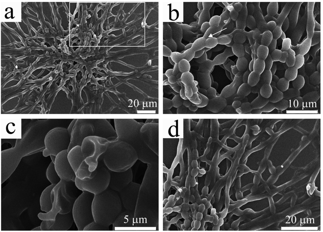
Figure 2.
SEM images of PVP aggregates formed at the early stage. (a) An overall view of an aggregate with a core (made of pearl-necklace-like linear chains) from which all the fibers were grown; (b) A closer view of the core from the aggregate shown in (a), highlighting that the formation and fusion of PVP microspheres into pearl-necklace-like chains (It should be noted that the branched structures are found in the core, as indicated by the arrow); (c) The pearl-necklace-like chains with some broken microspheres highlighting the hollow nature of PVP microspheres; d) A closer view of the area highlighted by a white square highlighted in (a), showing that free PVP molecules in the aqueous solutions continued to assemble onto the microspheres at the end of the pearl-necklace-like chains, and finally resulted in the formation of jellyfish-like large aggregate.

