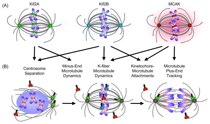
Fig. 1.
Localization and function of Kinesin-13s in mitosis. (A) The vertebrate Kinesin-13s have overlapping localizations at spindle poles and kinetochores/centromeres. MCAK is also found in the cytoplasm and at the plus-ends of microtubules. Kif2A localizations are shown in green, Kif2B in teal, and MCAK in red. Microtubules are shown in gray and chromosomes are shown in blue. (B) Summary of vertebrate Kinesin-13 function during mitosis. Spindle structures and Kinesin-13 models are colored as in (A) with kinetochores depicted in pink. Models of the Kinesin-13s are shown at their predominant site(s) of function in prophase, metaphase, and anaphase.

