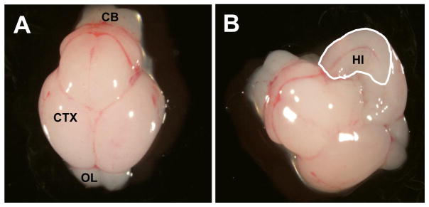
Figure 1.
Photographs of the dorsal view of the gestational day 17 murine brain. (A) The intact fetal brain on gestational day 17. (B) A unilateral view of the interior portion of the fetal cerebral cortex on gestational day 17. The cerebral cortex was opened along the midline and the hippocampal region was identified, removed and frozen on dry ice. The region demarcated by the white line represents the hippocampal tissue that was bilaterally excised from the cerebral cortex of the gestational day 17 murine fetus for RNA isolation. (CB) Cerebellum; (CTX) Cerebral cortex/Cerebrum; (OL) Olfactory lobes; (HI) Hippocampus.

