Fig. 4.
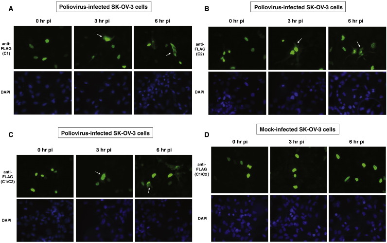
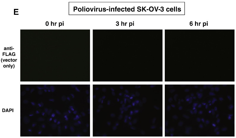
Sub-cellular localization of FLAG-tagged hnRNP C proteins transiently expressed in SK-OV-3 cells infected with poliovirus or mock-infected. Cells were processed and examined by fluorescence microscopy using a FITC filter at 0, 3, and 6 h post-infection. Green fluorescence indicates localization of FLAG-tagged hnRNP C1 in poliovirus-infected cells (A), FLAG-tagged hnRNP C2 in poliovirus-infected cells (B), FLAG-tagged hnRNP C1 and C2 in poliovirus-infected cells (C), and FLAG-tagged hnRNP C1 and C2 in mock-infected cells (D). Background fluorescence in cells transfected with empty vector pcDNA 3.1 is seen in (E). All cells were counterstained with DAPI to visualize nuclei.


