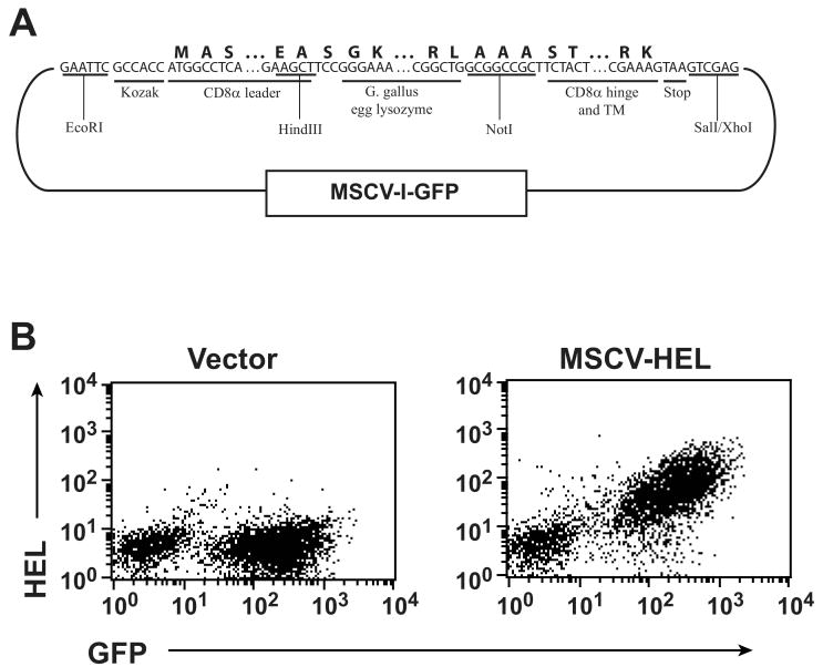
Figure 1. HEL expression on CTL.
(A) Diagram of the retroviral vector used to express HEL on CTL. Nucleotide sequence shows junctional segments between the chimeric gene's components. Amino acid sequence is shown above this and restriction sites used for cloning and segment identification below. Additional sequence can be found under the following GenBank accessions: G gallus lysozyme, NM_205281; CD8, XM_132621. (B) Retrovirus containing the MSCV-I-GFP vector or the HEL construct shown was used to transduce activated, purified CD8 T cells. Cells were surface stained with a HEL-specific antibody and flow cytometrically analyzed 5 days after transduction.

