
Figure 5. Co-transfer of MD-4 B cells and modified CTL into wild type mice.
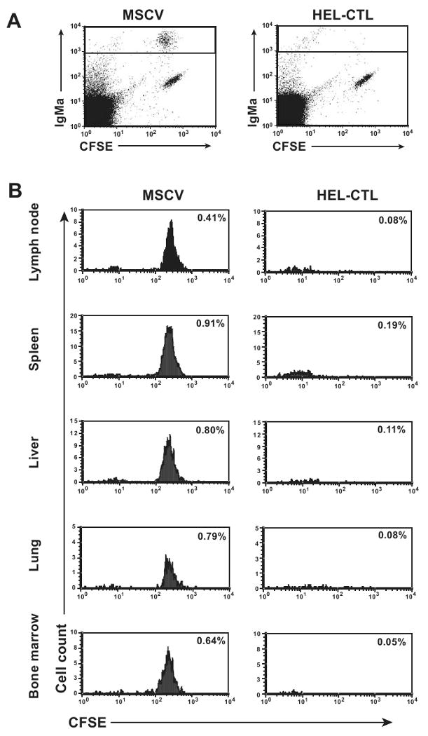
CFSE-labeled MD-4 splenocytes were adoptively transferred into wild-type C57BL/6 mice i.v. retro-orbitally and HEL- or control vector-modified CTL then transferred through the alternate retro-orbital plexus. 4 days later, the indicated organs were isolated and lymphocytes purified. Cells were stained for IgMa and analyzed by flow cytometry. (A) Representative dot plots of LN cells, gated for lymphocytes based on scatter properties, showing IgMa versus CFSE fluorescence. (B) Histogram plots of CFSE-staining among IgMa+ lymphocytes, gated based on the box shown in (A), is shown. Plots are normalized so that an equal number of scatter-gated lymphocytes were analyzed for the paired HEL-CTL and vector control plots to allow visual comparability. Percent of total lymphocytes that are IgMa+ MD-4 B cells is indicated. Representative plots are shown.