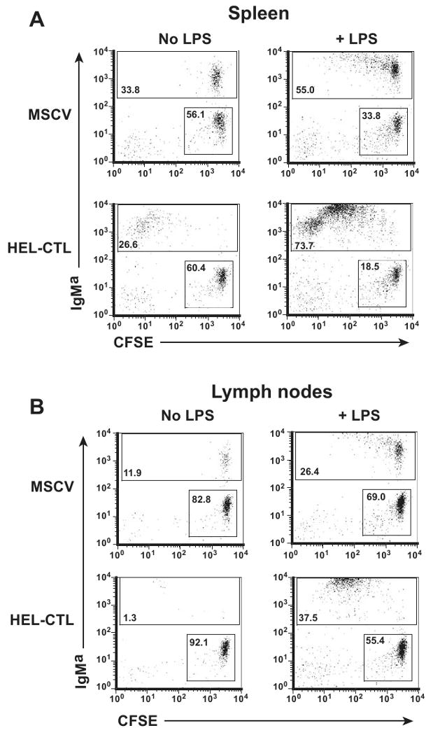
Figure 6. LPS promotes the survival of proliferating, HEL-CTL-treated MD-4 B cells.

MD-4 mice were bred onto the CD45.1 background, and CFSE-labeled splenocytes transferred into congenic C57BL/6 (CD45.1-CD45.2+) mice. HEL-CTL or vector-control CTL were transferred as in figure 5. LPS or saline (No LPS) was administered i.p. on the day of transfer. Lymphocytes in the spleen and LN were isolated 6 d later, and gated on the adoptively transferred CD45.1+ cells. (A) IgMa and CFSE staining among splenocytes. Numbers of dots on the different plots were normalized to show approximately equivalent numbers of IgMa- CFSE+ cells, which comprise primarily transferred T cells. Percent of IgMa- CFSE+ and IgMa+ cells are indicated in the respective boxes. (B) Analyses were performed as in (A), but for LN cells. Representative plots are shown.
