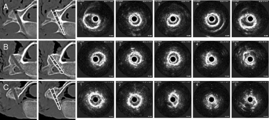
Fig. 2.

CT scans and corresponding IOUS images of experimental pedicle screw holes in the cervical spine. The numbers on the CT images in the second column show the position in which the ultrasound probe was situated when the IOUS images with the corresponding number on the right hand side were taken. a Shows a correctly placed trajectory. All intra-osseous images (right, 1-5) show an intact ring-like structure, representing the luminal surface of the pedicle screw hole. b Shows a slightly deviated pedicle screw hole. Note the deformed luminal echo in 4 and the partial loss of the highly intense ring-like structure in 5, corresponding to the perforation into the bony path of the vertebral artery. c Shows a completely deviated pedicle screw hole. While IOUS images 1 and 2 show regular appearances of an intra-osseous pedicle screw hole, a deformed signal is already seen in 3, while 4 and 5 show complete loss of the circular shape of the high-intensity bone signal and replacement of the low-intensity signal of the lumen of the pedicle screw hole by soft tissue reflectivity close to the probe
