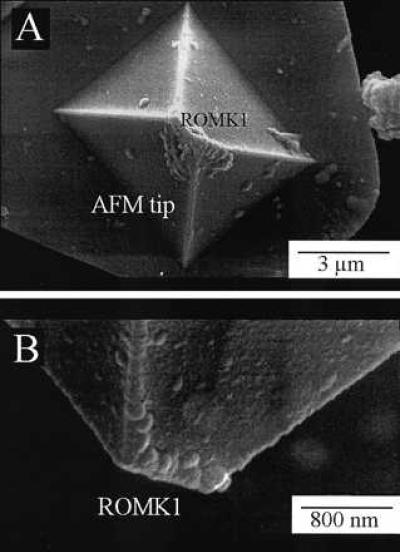
Figure 2.

Top view (A) and side view (B) of an AFM tip coated with the ROMK1 protein. Images were obtained with a scanning electron microscope (DSM 962, Zeiss). AFM tips were directly sputtered with 20- to 30-nm gold and then analyzed by scanning electron microscope. Note the bar-shaped AFM tip with ROMK1 proteins attached to it.
